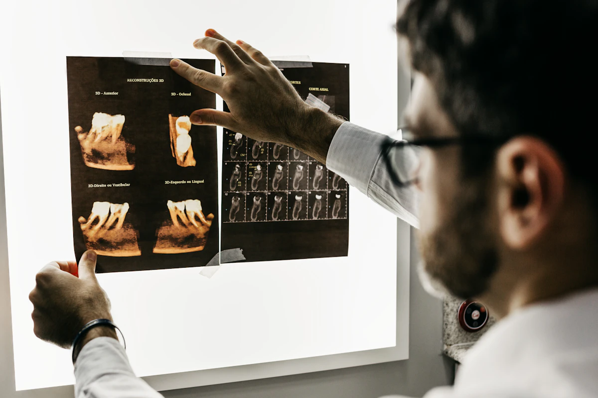
Understanding Your Bone X-Ray Report: Fractures, Osteoporosis, and Joints
Confused by terms like osteopenia, subluxation, or hairline fracture on your bone X-ray report? Learn what orthopedic radiologists look for and when to act.

Short answer: A bone X-ray report details bone alignment, cortical integrity, joint spacing, and bone density. Key terms like hairline fracture mean a non-displaced crack, osteopenia/osteoporosis indicate reduced bone mineral density, and joint space narrowing reflects cartilage wear from osteoarthritis.
You slipped on a patch of ice, twisted your ankle playing soccer, or finally decided to investigate persistent, aching pain in your knee or hip. After a visit to the urgent care clinic or imaging center, you receive your radiology results. Instead of a straightforward “it’s broken” or “it’s fine,” your document is filled with cryptic diagnostic phrases: “no discrete cortical breach,” “moderate joint space narrowing with marginal osteophyte formation,” or “diffuse demineralization with mild subluxation.”
When searching for a clear bone X-ray report explained, patients often find themselves caught between dense orthopedic textbooks and confusing online forums. The technical terminology used by musculoskeletal radiologists is designed to communicate precise anatomical realities to orthopedic surgeons and primary care doctors, but it can sound alarming to anyone else.
In this guide, we break down how radiologists systematically evaluate your bones, translate the most common radiographic terms into plain English, explain why early stress fractures are often invisible, and highlight critical emergency red flags.
The 4 Core Elements Radiologists Examine on Every Bone X-Ray
Musculoskeletal radiologists follow an organized diagnostic routine when reviewing any plain radiograph of your arms, legs, spine, or joints. They systematically assess four distinct structural domains:
- Alignment: How the bones sit relative to one another. Radiologists check for normal anatomical alignment versus subluxation (partial slippage of a joint) or dislocation (complete separation of joint surfaces).
- Cortical Margins and Bone Architecture: The dense outer shell of a bone is called the cortex. Radiologists inspect every millimeter of the cortex for cortical disruption (a step-off, buckle, or sharp lucent line indicating a fracture), as well as lytic (bone loss/holes) or sclerotic (abnormally dense/hardened) areas.
- Cartilage and Joint Spaces: Cartilage itself contains water and soft proteins, so it is transparent on an X-ray and appears as an open gap between bones. Radiologists evaluate whether this gap is symmetrical and healthy or narrowed by chronic friction and wear.
- Soft Tissues: While X-rays are calibrated for bones, surrounding soft tissues still show up in subtle shades of gray. Doctors look for joint effusion (excess fluid bulging the joint capsule), displaced fat pads (such as the “sail sign” in the elbow, which alerts doctors to a hidden fracture), and foreign bodies.
Decoding Common Bone Radiology Jargon: Plain-English Translations
When reviewing your imaging results, you will encounter specific descriptive phrases. Here is what they actually mean in everyday clinical practice:
| Radiographic Finding | Plain-English Translation | Most Common Clinical Causes | Typical Next Medical Step |
|---|---|---|---|
| “No acute fracture / Cortices intact” | Completely intact bone; no breaks, cracks, or structural disruption | Healthy skeleton, sprain, muscular strain, or contusion | Rest, ice, elevation (RICE protocol); physical therapy if pain persists |
| Hairline / Non-displaced Fracture | The bone is cracked all the way through, but the pieces remain perfectly aligned | Sports injuries, mild falls, stress accumulation | Immobilization with a splint, cast, or walking boot; protected weight-bearing |
| Displaced / Comminuted Fracture | Bone fragments have shifted out of alignment, or bone has shattered into 3+ pieces | High-impact trauma, motor vehicle collisions, falls from height | Urgent orthopedic reduction; often requires surgical fixation (plates/screws) |
| Joint Space Narrowing | The protective cartilage cushion between bones has thinned down significantly | Osteoarthritis (degenerative wear-and-tear), post-traumatic arthritis | Low-impact exercise, physical therapy, NSAIDs, joint lubricating injections |
| Osteophytes (Bone Spurs) | Smooth bony projections growing along joint margins | The body’s natural attempt to stabilize an arthritic, rubbing joint | Weight management, targeted strengthening; surgical removal is rarely needed |
| Subchondral Sclerosis | Thickened, hardened bone appearing bright white right beneath the joint cartilage | Bone reacting to excess mechanical friction as cartilage wears away | Core feature of osteoarthritis; physical therapy and joint preservation therapy |
| Osteopenia / Demineralization | Bones appear thinner or more washed-out (darker/radiolucent) than expected | Natural aging, post-menopausal hormonal changes, early osteoporosis | DEXA bone density scan, blood calcium/vitamin D3 tests, resistance training |
The “Hidden Fracture” Phenomenon: Why Initial X-Rays Can Miss Hairline Cracks
One of the most perplexing experiences for patients is being told their initial X-ray was “clear,” only to be told two weeks later that they have a fracture.
How can a bone X-ray miss a broken bone?
- Early Hairline & Stress Fractures: Microscopic cracks—especially in the scaphoid bone of the wrist, the neck of the femur, or the metatarsals of the foot—can be thinner than the resolution limit of standard X-ray beams on day one.
- The Bone Resorption Lag: During the first 7 to 14 days of bone healing, specialized cells called osteoclasts travel to the injury site to clean away dead microscopic bone fragments. This cleaning process slightly widens the fracture line, making it visible as a distinct dark lucency on follow-up imaging.
- Periosteal Reaction (Callus Formation): By weeks two and three, osteoblasts begin depositing soft mineralized matrix (callus), creating a visible hazy white “bump” of healing bone on follow-up X-rays.
Clinical Rule of Thumb: If you have severe, pinpoint bony tenderness after an injury, but your first X-ray is reported as “unremarkable,” your physician will often treat you with a temporary splint and order a repeat X-ray in 10 to 14 days or request an MRI for definitive soft-tissue and occult bone marrow evaluation.
Osteoarthritis vs. Osteoporosis on X-Ray: What Is the Difference?
Patients frequently confuse these two common bone conditions because both start with “osteo-” (meaning bone). On an X-ray, they look fundamentally different:
- Osteoarthritis (OA) is a Joint Disease:
- Affects articulating surfaces where two bones meet (knees, hips, fingers, spine).
- Hallmark X-ray triad: asymmetrical joint space narrowing, subchondral sclerosis (bright white bone beneath the joint), and marginal osteophytes (bone spurs).
- The bone substance itself is usually dense and strong; the joint surface is what has worn down.
- Osteoporosis is a Systemic Bone Density Disease:
- Affects the entire skeleton, making the interior bone matrix porous and fragile.
- Hallmark X-ray appearance: cortical thinning (the outer bone shell looks like a thin pencil line) and increased radiolucency (bones look dark or transparent instead of solid gray-white).
- Standard plain X-rays cannot reliably diagnose mild bone loss; an X-ray only reveals osteopenia after approximately 30% to 40% of bone mass has already been depleted. A specialized DEXA scan is required for formal T-score diagnosis.
When a Bone X-Ray Finding Demands Immediate Emergency Care
Most bone X-ray findings represent outpatient conditions that heal with casting, rest, or joint therapy. However, specific radiographic observations indicate an orthopedic emergency:
Emergency Red Flags: Seek immediate emergency medical care (call 911 or head to the nearest emergency trauma center) if your report or physical symptoms involve:
- Open (Compound) Fracture: Any fracture associated with an open skin wound or visible bone protruding through the skin (extreme risk of deep bacterial bone infection / osteomyelitis).
- Neurovascular Compromise: Numbness, tingling, severe pallor, cold skin, or loss of pulses in the fingers or toes beyond the fractured limb (indicates blood vessel or nerve entrapment).
- Unstable Pelvic or Spinal Fracture: Severe structural disruption of the pelvic ring or vertebral column carrying immediate risk of massive internal bleeding or spinal cord injury.
- Complete Joint Dislocation: Complete displacement of the hip, shoulder, elbow, or knee joint surfaces requiring urgent closed reduction to restore vital microcirculation.
- Aggressive Bone Destruction (Lytic Lesion / Sunburst Pattern): Irregular bone destruction with cortical breakthrough or a “sunburst” periosteal reaction, requiring urgent oncology evaluation to rule out primary bone sarcoma or metastatic disease.
4 Practical Steps for Reviewing Your Bone X-Ray Report
- Focus on the “Impression” first: The impression at the conclusion of the report summarizes the radiologist’s definitive clinical verdict. If it says “No acute osseous abnormality,” your bones have sustained no visible fracture.
- Verify weight-bearing instructions: If your report notes a “nondisplaced fracture” or “stress response,” do not walk on that limb until your treating physician confirms whether you are Non-Weight Bearing (NWB), Partial Weight Bearing (PWB), or Weight Bearing as Tolerated (WBAT).
- Remember that X-rays do not show soft tissue tears: If you heard a “pop” in your knee or ankle and cannot bear weight, a normal X-ray only rules out a broken bone—it does not rule out a torn ACL, meniscus, or Achilles tendon.
- Follow up in 10 to 14 days if pain persists: If focal bone tenderness continues despite a negative initial X-ray, schedule an orthopedic follow-up for repeat films or an MRI.
Questions to Ask Your Doctor or Orthopedic Specialist
- Does the fracture line extend into the joint surface (intra-articular fracture), and does that increase my risk of post-traumatic arthritis?
- Is this fracture stable enough to be treated with a removable splint or cast, or does it require surgical pin or plate fixation?
- Are the degenerative changes noted (joint space narrowing, bone spurs) consistent with normal aging, or do they warrant physical therapy and joint injections?
- Should I schedule a DEXA bone density scan to evaluate for underlying osteoporosis, especially if this fracture occurred from a simple, low-energy fall?
Summary
A bone X-ray report is an objective structural assessment of your skeletal framework. Terms like osteopenia, cortical disruption, or joint space narrowing describe physical bone density and alignment. The ultimate treatment plan depends on correlating these radiographic images with your pain, mobility, and physical examination.
If you have received an orthopedic radiology report full of confusing jargon and want an instant, plain-English breakdown before your doctor’s appointment, MedGPT can help translate your findings and organize your clinical questions.
Want Plain-English Explanations of Your Bone X-Ray?
Upload or paste your bone X-ray report into the MedGPT app to receive clear, structured medical translations of orthopedic findings in seconds.
Download the MedGPT App
Medical Disclaimer: This article is intended solely for educational and informational purposes and does not substitute for professional medical advice, clinical diagnosis, or orthopedic radiological interpretation. Always discuss imaging results and treatment plans directly with your treating physician or orthopedic specialist.
Sources & Citations
MedGPT relies on internationally recognized medical organizations and research to ensure our content is accurate, trustworthy, and evidence-based.
- American Academy of Orthopaedic Surgeons (AAOS) - OrthoInfo Fractures GuideClinical guidelines on fracture classification, bone healing, and orthopedic radiological evaluation.
- Radiological Society of North America (RSNA) - Bone Radiography (X-ray)Patient overview of skeletal X-ray procedures, normal anatomy, and radiographic signs of joint disease.
- International Osteoporosis Foundation (IOF) - Diagnostic Imaging GuidelinesStandard protocols for identifying osteoporotic vertebral fractures and metabolic bone changes on plain radiography.