Dental X-Ray Report: Cavities, Root Canals & Jaw Health
Confused by terms like periapical radiolucency, interproximal caries, or bone loss on your dental X-ray? Learn what dentists look for and what it means.
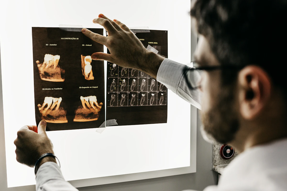
Short answer: A dental X-ray report evaluates teeth, roots, and jawbone density using minimal radiation. Dark areas (radiolucencies) signify decay, infection, or nerve spaces, while bright white structures (radiopacities) indicate enamel, bone, fillings, or crowns.
You settle into the dental chair for your routine checkup or an emergency visit for a throbbing molar. The hygienist places a small plastic sensor in your mouth, instructs you to bite down, and steps behind a protective shield for a few seconds. Moments later, high-resolution grayscale images illuminate the monitor above your chair: “Let’s take a look at your dental X-rays.”
Your dentist points to the screen, tracing shadows and white outlines with an explorer probe: “You have interproximal caries on number 19 entering the dentin,” “there is widening of the periodontal ligament space with a periapical radiolucency at the apex of number 30,” or “we are noticing generalized horizontal crestal bone loss consistent with moderate periodontitis.”
When patients search for a dental X-ray report explained, they are often caught between two extremes: confusion over why a tooth that feels completely fine needs an expensive root canal or filling, or anxiety that dark spots on their jawbone represent tumors.
In this comprehensive clinical guide, we demystify how dentists and oral maxillofacial radiologists read dental radiographs, translate confusing radiographic jargon into plain English, review radiation safety, and highlight urgent dental red flags that require immediate intervention.
How Dental X-Rays Work: Radiolucency vs. Radiopacity
Dental radiography relies on controlled bursts of low-dose X-ray beams directed through oral tissues onto a digital phosphor plate or solid-state electronic sensor. As X-rays pass through your mouth, different tissues absorb the beam at varying rates depending on their physical density:
- Radiolucent (Dark Gray to Pitch Black): Structures that are hollow, soft, or demineralized offer little resistance to X-rays. The beams pass straight through and strike the sensor, creating dark areas on the image. Normal radiolucencies include the dental pulp chamber, root canals, and the periodontal ligament space. Pathological radiolucencies include active cavities, bacterial demineralization, cysts, and root-tip abscesses.
- Radiopaque (Light Gray to Brilliant White): Denser mineralized tissues absorb the majority of the X-ray beam, blocking it from reaching the digital sensor. Enamel (the hardest substance in the human body) appears as a bright white outline; dentin appears slightly grayer; cortical jawbone appears textured white. Dental materials like amalgam fillings, gold, porcelain-fused-to-metal crowns, titanium implants, and gutta-percha root canal fillings appear bright, glowing white.
[ Pitch Black ] ----------> [ Medium Gray ] ----------> [ Pure White ]
Pulp chamber, decay,         Dentin, spongy bone,        Enamel, fillings,
abscesses, bone loss         cementum                    crowns, implants
(Radiolucent)                                            (Radiopaque)
The 4 Primary Types of Dental X-Rays
Dentists use different radiographic views depending on whether they are checking for cavities between teeth, examining root health, or assessing overall facial anatomy:
- Bitewing Radiographs (BWX):
- What they show: The crowns of upper and lower posterior teeth (premolars and molars) biting together in a single view, alongside the crest of the supporting alveolar jawbone.
- Primary diagnostic role: The undisputed gold standard for detecting interproximal caries (cavities forming hidden between tight teeth contacts) and measuring early periodontal bone loss.
- Periapical Radiographs (PA):
- What they show: The entire tooth from the biting crown all the way to the tip of the root (the apex), including the surrounding jawbone.
- Primary diagnostic role: Investigating deep toothaches, detecting root fractures, assessing root canal morphology, and identifying periapical abscesses or granulomas at the root tip.
- Panoramic Radiograph (OPG / Panorex):
- What they show: A broad 2D panoramic sweep capturing both jaws, all teeth, the maxillary sinuses, and both temporomandibular joints (TMJs) in a single wide image.
- Primary diagnostic role: Evaluating wisdom teeth (third molar impaction), planning orthodontic treatment, assessing jawbone cysts or fractures, and screening bone health before full-mouth rehabilitation.
- Cone Beam Computed Tomography (CBCT):
- What they show: A 3D volumetric reconstruction of dental anatomy with microscopic resolution.
- Primary diagnostic role: Dental implant placement planning, navigating complex or calcified root canals in endodontics, and mapping roots located dangerously close to the mandibular nerve canal.
Anatomy of a Tooth on a Radiograph
To understand what is wrong on a dental X-ray, you must first recognize normal anatomy:
      Crown --->  [ Enamel (Dense White)       ]
                  [ Dentin (Mid-Gray Core)      ]
                  [ Pulp Chamber (Dark Center)  ]
       Neck --->  [ Cementoenamel Junction (CEJ)]
       Root --->  [ Periodontal Ligament (PDL)  ] <--- Thin dark line
                  [ Lamina Dura (Socket Wall)   ] <--- Thin white line
Apex ---> [ Alveolar Bone (Trabecular) ]- Enamel & Dentin: The protective mineralized shell of the tooth crown.
- Pulp Chamber & Canals: The living center of the tooth housing nerves and blood vessels. In young patients, pulp chambers are wide; as we age or experience wear, the pulp chamber naturally calcifies and shrinks.
- Lamina Dura: A crisp, continuous white line that forms the inner bony socket enclosing the root. Disruption or loss of the lamina dura is one of the earliest signs of active infection or occlusal trauma.
- Periodontal Ligament (PDL) Space: A uniform, paper-thin dark ribbon separating the tooth root from the white lamina dura. Widening of this space signals acute inflammation, tooth concussion, or biting overload.
- Alveolar Crest: The top margin of the supporting jawbone. Normally, bone sits roughly 1.5 to 2.0 mm below the cementoenamel junction (where crown meets root).
Decoding Common Dental X-Ray Findings
| Radiographic Finding | What It Actually Means in Plain English | Most Common Clinical Cause | Typical Next Dental Step |
|---|---|---|---|
| Interproximal Radiolucency in Enamel | Early chemical demineralization between teeth, confined to enamel | Bacterial plaque accumulation, infrequent flossing | Topical fluoride, remineralization therapy, monitor without drilling |
| Carious Radiolucency into Dentin | Active cavity has breached the enamel shield into the softer dentin | Ongoing acid erosion by cariogenic bacteria | Composite resin filling or ceramic inlay/onlay |
| Recurrent / Secondary Caries | Dark shadow forming at the edge or floor of an existing filling | Marginal breakdown of restoration, microleakage | Replacement of filling; crown if remaining tooth structure is compromised |
| Periapical Radiolucency (PARL) | Dark circular shadow surrounding the root apex | Necrotic (dead) nerve tissue, chronic apical abscess | Root canal therapy (endodontics) or tooth extraction |
| Horizontal Bone Loss | Alveolar jawbone level has dropped uniformly across multiple teeth | Chronic periodontitis, genetic predisposition, smoking | Deep scaling and root planing (SRP), periodontal maintenance cleanings |
| Vertical / Angular Bone Defect | Deep, V-shaped trench of bone loss isolated alongside one root | Advanced localized periodontitis, food impaction | Periodontal pocket debridement, bone grafting, regenerative therapy |
| Subgingival Calculus Spur | Tiny radiopaque “spurs” or “ledges” protruding from the root | Hardened mineralized tartar beneath the gumline | Scaling and root planing to prevent rapid periodontal detachment |
| Impacted Third Molar (Wisdom Tooth) | Wisdom tooth trapped beneath bone or gum, angled sideways | Inadequate jaw arch length, evolutionary jaw size reduction | Prophylactic surgical extraction if causing damage to second molar |
| Condylar Flattening / Sclerosis | Joint head of the lower jaw appears worn down or heavily calcified | Temporomandibular joint osteoarthritis, severe bruxism | Nightguard (occlusal splint), physical therapy, joint evaluation |
Cavities on X-Ray: Why Did My Dentist Say I Need a Filling When I Feel No Pain?
One of the most common questions patients ask is: “If my tooth doesn’t hurt, why does the X-ray show a cavity?”
Tooth enamel has zero nerve endings. In fact, a cavity can eat halfway through the protective dentin layer before a patient experiences any noticeable temperature sensitivity or twinges on sweets.
- Incipient (Enamel-Only) Caries: Appears as a faint, triangular radiolucent notch on the outer enamel. These lesions do not require drilling. With prescription high-fluoride toothpaste, xylitol, and disciplined interdental flossing, the mineral matrix can remineralize and arrest naturally.
- Moderate-to-Advanced (Dentin) Caries: Once bacterial acid penetrates past the dentinoenamel junction (DEJ), it spreads rapidly through microscopic dentinal tubules like an umbrella. At this stage, drilling away the bacteria and placing an adhesive composite restoration is necessary to protect the living pulp.
- Pulp-Involved Caries: The dark shadow extends directly into the central nerve chamber. At this stage, a simple filling is no longer sufficient; the tooth requires root canal treatment to prevent spreading infection.
Root Canals Explained: What Is a “Periapical Radiolucency”?
When a dental report notes a “periapical radiolucency” (PARL) or “apical rarefying osteitis,” it signifies that the nerve tissue inside the pulp has died (pulpal necrosis), usually from deep decay, trauma, or a cracked tooth.
Bacterial toxins leak out through the microscopic opening at the very tip of the root (the apical foramen) into the surrounding jawbone. Your body’s immune cells rush to the site, releasing chemical mediators that break down neighboring trabecular bone to make room for defense cells. On the X-ray, this bone destruction shows up as a dark circular halo around the root tip.
- Can a tooth with an abscess have zero pain? Yes! Chronic periapical abscesses or granulomas often develop silently over months. If the tooth nerve is dead, it cannot transmit sharp pain signals.
- The Solution: A root canal cleans out the infected hollow canals, disinfects them with antibacterial irrigants, and seals them with a biocompatible rubber material called gutta-percha (which appears radiopaque bright white on post-treatment X-rays).
Radiation Safety: How Safe Are Modern Dental X-Rays?
Many patients worry about cancer risk from routine dental imaging. However, thanks to ultra-fast digital sensors, rectangular collimation, and low-voltage generators, modern dental radiographs produce astonishingly low doses of radiation:
- 4 Digital Bitewing X-Rays: Approximately 0.005 millisieverts (5 µSv).
- Panoramic X-Ray (OPG): Approximately 0.010 to 0.020 mSv (10–20 µSv).
- For Comparison:
- Natural background radiation received from the environment, soil, and space: ~8 µSv every single day.
- A 4-hour commercial airline flight: ~20 µSv of cosmic radiation.
- Chest CT scan: ~7.0 mSv (7,000 µSv).
A standard set of dental bitewings exposes you to roughly the same radiation dose as spending an afternoon in the park or eating two bananas (due to naturally occurring potassium-40 isotopes). Denying diagnostic dental X-rays carries far greater health risks, as hidden bone loss and infections remain undetected until irreversible damage occurs.
Urgent Dental Red Flags
While most dental X-ray findings represent routine chronic issues, specific symptoms accompanying radiographic findings constitute medical and surgical emergencies:
Emergency Red Flags (Seek Immediate Medical or Dental Attention):
- Ludwig’s Angina: Rapidly expanding bilateral swelling of the submandibular and sublingual spaces following a lower molar abscess. Manifests as a raised tongue, difficulty swallowing (dysphagia), and airway compromise. This is a life-threatening emergency.
- Canine Space / Periorbital Cellulitis: Severe swelling spreading toward the cheek, eye socket, and bridge of the nose from an upper front tooth or premolar, carrying a risk of cavernous sinus thrombosis.
- Trismus with Spreading Facial Swelling: Inability to open the mouth wider than 15–20 mm accompanied by fever and jaw stiffness.
- Sudden Malocclusion After Facial Trauma: Teeth no longer fitting together properly, accompanied by pain on biting, indicating a fractured mandible (lower jaw) or maxilla.
4 Practical Steps for Reviewing Your Dental X-Rays
- Ask your dentist to zoom in on the tooth numbers: In the United States, teeth are numbered 1 through 32 (starting upper right third molar). Internationally, the two-digit FDI system is used (e.g., tooth 11 to 48). Make sure you know exactly which tooth is being discussed.
- Distinguish between active cavities and old restorations: Metallic fillings appear brilliant glowing white, while composite resin fillings may appear grayish-white. Ask if the dark shadow is beneath an old filling (recurrent decay) or between virgin teeth.
- Inquire if early lesions can be remineralized: If a cavity is strictly confined within the enamel layer on your bitewing, ask whether prescription 5,000 ppm fluoride toothpaste and rigorous hygiene can be attempted for 6 months before drilling.
- Compare crestal bone levels over time: Ask your dentist to overlay your current bitewings with those from 2 or 3 years ago to determine if your bone loss is active or stabilized.
Questions to Ask Your Dentist
- Is the dark shadow on this tooth confined solely to the enamel, or has it penetrated into the dentin?
- Does the periapical shadow around my root represent an active infection requiring endodontic therapy, or stable healing scar tissue from a prior procedure?
- What percentage of supporting jawbone has been lost around my molars, and is it horizontal or vertical?
- Is my impacted wisdom tooth causing pressure or external root resorption on the adjacent second molar?
Summary
A dental X-ray report is an essential window into the unseen 60% of your tooth structure and supporting jawbone hidden beneath the gums. Radiographic terms like interproximal radiolucency, periodontal ligament widening, or alveolar crestal resorption describe the mineral density of your teeth and bones rather than permanent disasters. Early detection allows for minimally invasive, tooth-preserving treatments long before toothaches occur.
If your dental clinic provided you with an X-ray report or digital radiographs and you want plain-English explanations of cavities, root canals, or bone levels before committing to treatment, MedGPT can help decode your dental imaging in seconds.
Want Plain-English Explanations of Your Dental X-Rays?
Upload or paste your dental radiographic report into the MedGPT app to receive clear, structured medical translations of cavities, root canals, and jawbone findings in seconds.
Download the MedGPT App
Medical Disclaimer: This article is intended solely for educational and informational purposes and does not substitute for professional dental advice, clinical diagnosis, or radiographic interpretation. Always discuss imaging results and treatment plans directly with your licensed dentist, periodontist, or oral surgeon.
Sources & Citations
MedGPT relies on internationally recognized medical organizations and research to ensure our content is accurate, trustworthy, and evidence-based.
- American Dental Association (ADA) - Dental Radiographic ExaminationsClinical guidelines on patient selection, radiation safety protocols, and diagnostic imaging efficacy in oral healthcare.
- European Federation of Periodontology (EFP)International consensus on radiographic assessment of alveolar bone levels and periodontal disease progression.
- American Association of Endodontists (AAE) - Endodontic RadiographyDiagnostic standards for evaluating periapical lesions, pulp vitality, and root canal obturation.